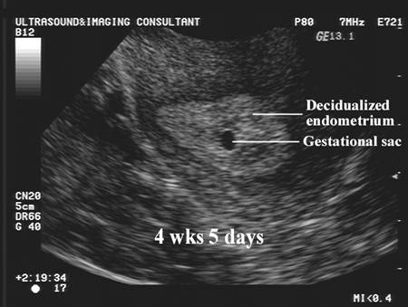
Gestational Sac 1 2 Weeks Pregnant Ultrasound Pictures
Diagnostic Ultrasound In The First Trimester Of Pregnancy Glowm
Diagnostic Ultrasound In The First Trimester Of Pregnancy Glowm
Gestational Sac During Pregnancy

Pin On Ob Gyn Ultrasound 101 Mod 2
Diagnostic Ultrasound In The First Trimester Of Pregnancy Glowm
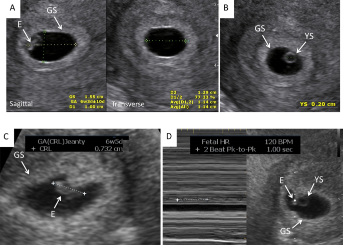
Early Pregnancy Ultrasound Measurements And Prediction Of First Trimester Pregnancy Loss A Logistic Model Scientific Reports

There s no ultrasound image of your baby to be for weeks 1 and 2.
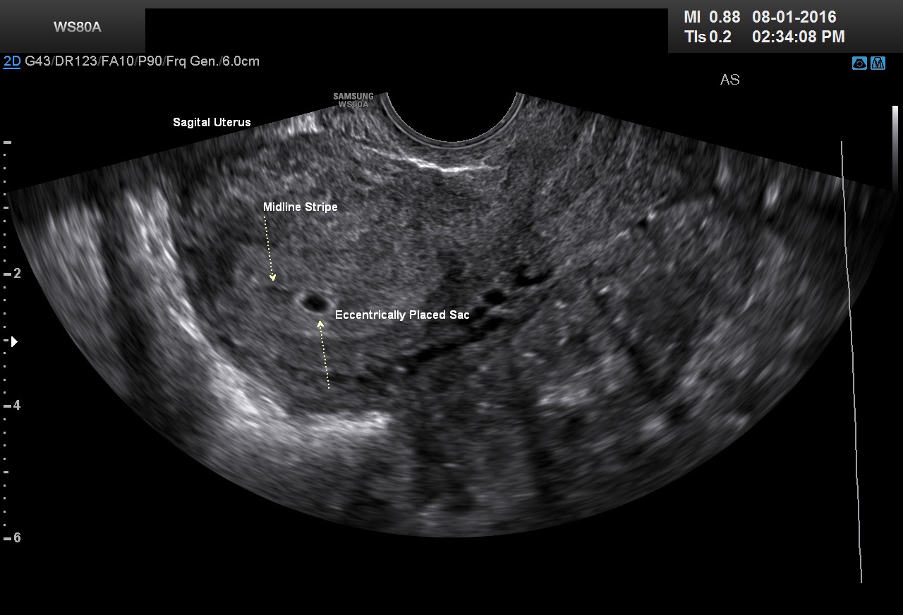
Gestational sac 1 2 weeks pregnant ultrasound pictures. Abnormalities in size and appearance of the yolk sac have been reported to be associated with an increased risk of pregnancy loss although not all studies have found this to be a useful metric for predicting pregnancy loss. Its normal eccentric location. This develops within the gestational sac and provides nutrition to the developing embryo. The yolk sac usually becomes visible on a transvaginal ultrasound between 5 1 2 and 6 weeks gestation.
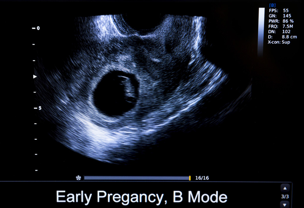
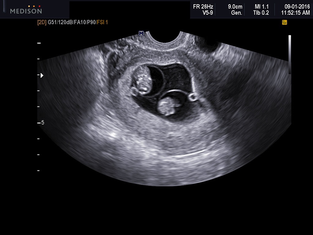
It can be seen via transvaginal ultrasound between 3 5 weeks pregnant. The fetal pole is the first direct imaging manifestation of the fetus and is seen as a thickening on the margin of the yolk sac during early pregnancy it is often used synonymously with the term embryo. The yolk sac is the first structure that is identifiable within the gestational sac by transvaginal ultrasound when the gestational sac reaches 8 10 mm. The gestational sac increases in diameter by 1 13 mm per day and initially measures 2 to 3 mm in diameter according.
First trimester ultrasound pictures. The next positive sign of pregnancy is seeing the yolk sac. In experienced hands it may be detected as early as 30 days gestation by tvu. A 2yr old maltese terrier 46 days post mating.
But that little sac is a kind of baby cocoon called a gestational sac. The fetal pole is usually identified at 6 5 weeks with transabdominal ultrasound imaging and at 6 weeks 2 with transvaginal ultrasound imaging although it may not be seen until 9 weeks in. Right lateral abdominal radiograph of 2yr old poodle x 30d post ai with no puppies visible. A true gestational sac can be distinguished from a pseudogestational sac by noting.
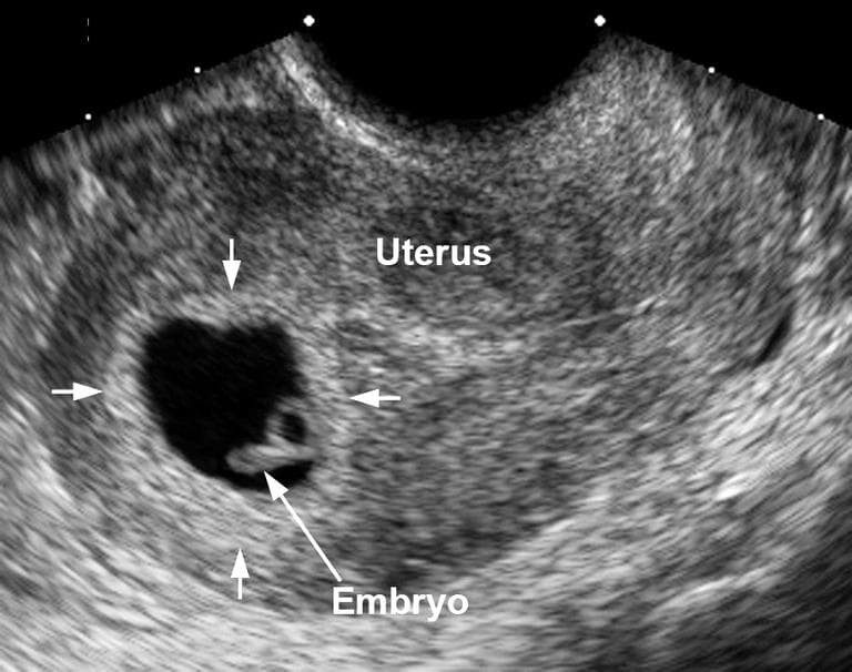
It is embedded in endometrium rather. You may see the gestational sac in an ultrasound as early as 4 1 2 to 5 weeks. The gestational sac is a structure that surrounds an embryo which is a baby in the very early stages of development it encloses not only the embryo but also the amniotic fluid which helps to nourish and protect the developing baby the gestational sac is the structure ultrasound technicians look for when they need to confirm the presence and viability of an early pregnancy. It initially appears as a round anechoic structure.
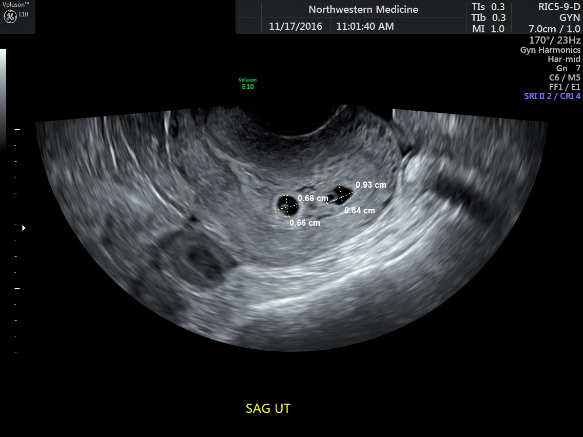
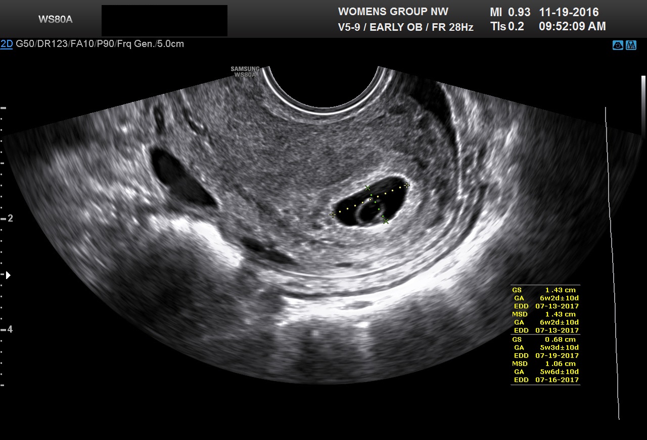
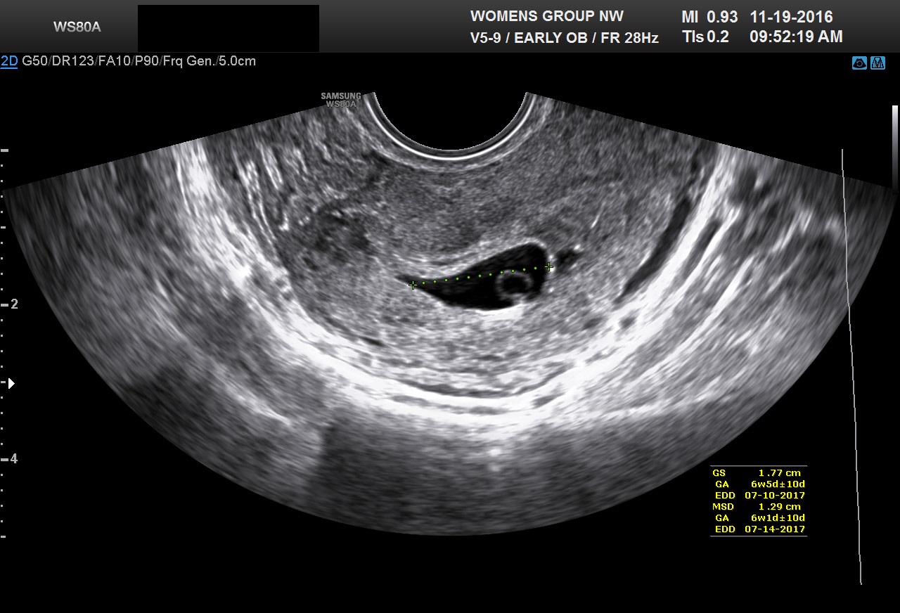
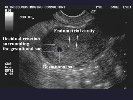
This can be seen on ultrasound by 5 1 2 to 6 weeks gestation. The gestational sac gs is the first sign of early pregnancy on ultrasound and can be seen with endovaginal ultrasound at approximately 3 5 weeks gestation when the mean sac diameter msd would approximately measure 2 3 mm in diameter.

4 Weeks Pregnant On Ultrasound The Early Gestational Sac Womb With A View Blog

Figure 1 From Ultrasound Evaluation Of The First Trimester Semantic Scholar
Diagnostic Ultrasound In The First Trimester Of Pregnancy Glowm
The Gestational Sac In Pregnancy Babymed Com

Small Gestational Sac On Ultrasound Ultrasound Ultrasound Pictures Baby Ultrasound
Tragically Wrong When Good Early Pregnancies Are Misdiagnosed As Bad Commonhealth

Pin On Ob Gyn Ultrasound 101 Mod 2
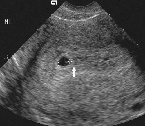
The Normal Gestational Sac

Patience Is Key Understanding The Timing Of Early Ultrasounds Your Pregnancy Matters Ut Southwestern Medical Center

First Trimester Ultrasound Early Pregnancy Failure Radiology Key
The Normal Gestational Sac
Diagnostic Ultrasound In The First Trimester Of Pregnancy Glowm
Obstetric And Gynecologic Imaging Radiology Key
/doctor-using-ultrasound-on-pregnant-woman-591405835-595195b43df78cae81c346ba.jpg)